Target Information
| Target General Information | Top | |||||
|---|---|---|---|---|---|---|
| Target ID |
T00348
|
|||||
| Target Name |
ABL T315I mutant (ABL T315I)
|
|||||
| Synonyms |
p150 T315I; T315I Abl; Proto-oncogene tyrosine-protein kinase ABL1 T315I; Proto-oncogene c-Abl T315I; JTK7 T315I; C-ABL T315I; Abl T315I; Abelson tyrosine-protein kinase 1 T315I; Abelson murine leukemia viral oncogene homolog 1 T315I
Click to Show/Hide
|
|||||
| Gene Name |
ABL1
|
|||||
| Target Type |
Patented-recorded target
|
[1] | ||||
| Function |
Coordinates actin remodeling through tyrosine phosphorylation of proteins controlling cytoskeleton dynamics like WASF3 (involved in branch formation); ANXA1 (involved in membrane anchoring); DBN1, DBNL, CTTN, RAPH1 and ENAH (involved in signaling); or MAPT and PXN (microtubule-binding proteins). Phosphorylation of WASF3 is critical for the stimulation of lamellipodia formation and cell migration. Involved in the regulation of cell adhesion and motility through phosphorylation of key regulators of these processes such as BCAR1, CRK, CRKL, DOK1, EFS or NEDD9. Phosphorylates multiple receptor tyrosine kinases and more particularly promotes endocytosis of EGFR, facilitates the formation of neuromuscular synapses through MUSK, inhibits PDGFRB-mediated chemotaxis and modulates the endocytosis of activated B-cell receptor complexes. Other substrates which are involved in endocytosis regulation are the caveolin (CAV1) and RIN1. Moreover, ABL1 regulates the CBL family of ubiquitin ligases that drive receptor down-regulation and actin remodeling. Phosphorylation of CBL leads to increased EGFR stability. Involved in late-stage autophagy by regulating positively the trafficking and function of lysosomal components. ABL1 targets to mitochondria in response to oxidative stress and thereby mediates mitochondrial dysfunction and cell death. In response to oxidative stress, phosphorylates serine/threonine kinase PRKD2 at 'Tyr-717'. ABL1 is also translocated in the nucleus where it has DNA-binding activity and is involved in DNA-damage response and apoptosis. Many substrates are known mediators of DNA repair: DDB1, DDB2, ERCC3, ERCC6, RAD9A, RAD51, RAD52 or WRN. Activates the proapoptotic pathway when the DNA damage is too severe to be repaired. Phosphorylates TP73, a primary regulator for this type of damage-induced apoptosis. Phosphorylates the caspase CASP9 on 'Tyr-153' and regulates its processing in the apoptotic response to DNA damage. Phosphorylates PSMA7 that leads to an inhibition of proteasomal activity and cell cycle transition blocks. ABL1 acts also as a regulator of multiple pathological signaling cascades during infection. Several known tyrosine-phosphorylated microbial proteins have been identified as ABL1 substrates. This is the case of A36R of Vaccinia virus, Tir (translocated intimin receptor) of pathogenic E. coli and possibly Citrobacter, CagA (cytotoxin-associated gene A) of H. pylori, or AnkA (ankyrin repeat-containing protein A) of A. phagocytophilum. Pathogens can highjack ABL1 kinase signaling to reorganize the host actin cytoskeleton for multiple purposes, like facilitating intracellular movement and host cell exit. Finally, functions as its own regulator through autocatalytic activity as well as through phosphorylation of its inhibitor, ABI1. Regulates T-cell differentiation in a TBX21-dependent manner. Phosphorylates TBX21 on tyrosine residues leading to an enhancement of its transcriptional activator activity. Non-receptor tyrosine-protein kinase that plays a role in many key processes linked to cell growth and survival such as cytoskeleton remodeling in response to extracellular stimuli, cell motility and adhesion, receptor endocytosis, autophagy, DNA damage response and apoptosis.
Click to Show/Hide
|
|||||
| BioChemical Class |
Kinase
|
|||||
| UniProt ID | ||||||
| EC Number |
EC 2.7.10.2
|
|||||
| Sequence |
MLEICLKLVGCKSKKGLSSSSSCYLEEALQRPVASDFEPQGLSEAARWNSKENLLAGPSE
NDPNLFVALYDFVASGDNTLSITKGEKLRVLGYNHNGEWCEAQTKNGQGWVPSNYITPVN SLEKHSWYHGPVSRNAAEYLLSSGINGSFLVRESESSPGQRSISLRYEGRVYHYRINTAS DGKLYVSSESRFNTLAELVHHHSTVADGLITTLHYPAPKRNKPTVYGVSPNYDKWEMERT DITMKHKLGGGQYGEVYEGVWKKYSLTVAVKTLKEDTMEVEEFLKEAAVMKEIKHPNLVQ LLGVCTREPPFYIITEFMTYGNLLDYLRECNRQEVNAVVLLYMATQISSAMEYLEKKNFI HRDLAARNCLVGENHLVKVADFGLSRLMTGDTYTAHAGAKFPIKWTAPESLAYNKFSIKS DVWAFGVLLWEIATYGMSPYPGIDLSQVYELLEKDYRMERPEGCPEKVYELMRACWQWNP SDRPSFAEIHQAFETMFQESSISDEVEKELGKQGVRGAVSTLLQAPELPTKTRTSRRAAE HRDTTDVPEMPHSKGQGESDPLDHEPAVSPLLPRKERGPPEGGLNEDERLLPKDKKTNLF SALIKKKKKTAPTPPKRSSSFREMDGQPERRGAGEEEGRDISNGALAFTPLDTADPAKSP KPSNGAGVPNGALRESGGSGFRSPHLWKKSSTLTSSRLATGEEEGGGSSSKRFLRSCSAS CVPHGAKDTEWRSVTLPRDLQSTGRQFDSSTFGGHKSEKPALPRKRAGENRSDQVTRGTV TPPPRLVKKNEEAADEVFKDIMESSPGSSPPNLTPKPLRRQVTVAPASGLPHKEEAGKGS ALGTPAAAEPVTPTSKAGSGAPGGTSKGPAEESRVRRHKHSSESPGRDKGKLSRLKPAPP PPPAASAGKAGGKPSQSPSQEAAGEAVLGAKTKATSLVDAVNSDAAKPSQPGEGLKKPVL PATPKPQSAKPSGTPISPAPVPSTLPSASSALAGDQPSSTAFIPLISTRVSLRKTRQPPE RIASGAITKGVVLDSTEALCLAISRNSEQMASHSAVLEAGKNLYTFCVSYVDSIQQMRNK FAFREAINKLENNLRELQICPATAGSGPAATQDFSKLLSSVKEISDIVQR Click to Show/Hide
|
|||||
| Drug Binding Sites of Target | Top | |||||
|---|---|---|---|---|---|---|
| Ligand Name: Axitinib | Ligand Info | |||||
| Structure Description | The crystal structure of human abl1 T315I gatekeeper mutant kinase domain in complex with axitinib | PDB:4TWP | ||||
| Method | X-ray diffraction | Resolution | 2.40 Å | Mutation | Yes | [2] |
| PDB Sequence |
DKWEMERTDI
242 TMKHKLGGGQ252 YGEVYEGVWK262 KYSLTVAVKT272 LEVEEFLKEA287 AVMKEIKHPN 297 LVQLLGVCTR307 EPPFYIIIEF317 MTYGNLLDYL327 RECNRQEVNA337 VVLLYMATQI 347 SSAMEYLEKK357 NFIHRDLAAR367 NCLVGENHLV377 KVADFGLSRL387 MTGDTYTAHA 397 GAKFPIKWTA407 PESLAYNKFS417 IKSDVWAFGV427 LLWEIATYGM437 SPYPGIDLSQ 447 VYELLEKDYR457 MERPEGCPEK467 VYELMRACWQ477 WNPSDRPSFA487 EIHQAFETMF 497 QESSIS
|
|||||
|
|
LEU248
3.616
GLY249
4.119
TYR253
2.880
VAL256
3.926
ALA269
3.644
LYS271
2.688
GLU286
4.062
VAL299
4.712
ILE315
4.301
GLU316
2.720
PHE317
3.332
|
|||||
| Ligand Name: PHA-739358 | Ligand Info | |||||
| Structure Description | Crystal structure of the T315I Abl mutant in complex with the inhibitor PHA-739358 | PDB:2V7A | ||||
| Method | X-ray diffraction | Resolution | 2.50 Å | Mutation | Yes | [3] |
| PDB Sequence |
DKWEMERTDI
242 TMKHKLGGGQ252 YGEVYEGVWK262 KYSLTVAVKT272 LKEDTMEVEE282 FLKEAAVMKE 292 IKHPNLVQLL302 GVCTREPPFY312 IIIEFMTYGN322 LLDYLRECNR332 QEVNAVVLLY 342 MATQISSAME352 YLEKKNFIHR362 DLAARNCLVG372 ENHLVKVADF382 GLSRLMTGDT 392 TAHAGAKFPI403 KWTAPESLAY413 NKFSIKSDVW423 AFGVLLWEIA433 TYGMSPYPGI 443 DLSQVYELLE453 KDYRMERPEG463 CPEKVYELMR473 ACWQWNPSDR483 PSFAEIHQAF 493 ETMFQESSIS503
|
|||||
|
|
LEU248
3.388
GLY250
3.174
GLY251
4.300
GLN252
4.946
VAL256
4.203
ALA269
3.405
LYS271
2.994
GLU286
4.523
VAL299
4.382
ILE315
4.125
GLU316
2.826
PHE317
3.681
|
|||||
| Click to View More Binding Site Information of This Target with Different Ligands | ||||||
| Different Human System Profiles of Target | Top |
|---|---|
|
Human Similarity Proteins
of target is determined by comparing the sequence similarity of all human proteins with the target based on BLAST. The similarity proteins for a target are defined as the proteins with E-value < 0.005 and outside the protein families of the target.
A target that has fewer human similarity proteins outside its family is commonly regarded to possess a greater capacity to avoid undesired interactions and thus increase the possibility of finding successful drugs
(Brief Bioinform, 21: 649-662, 2020).
Human Pathway Affiliation
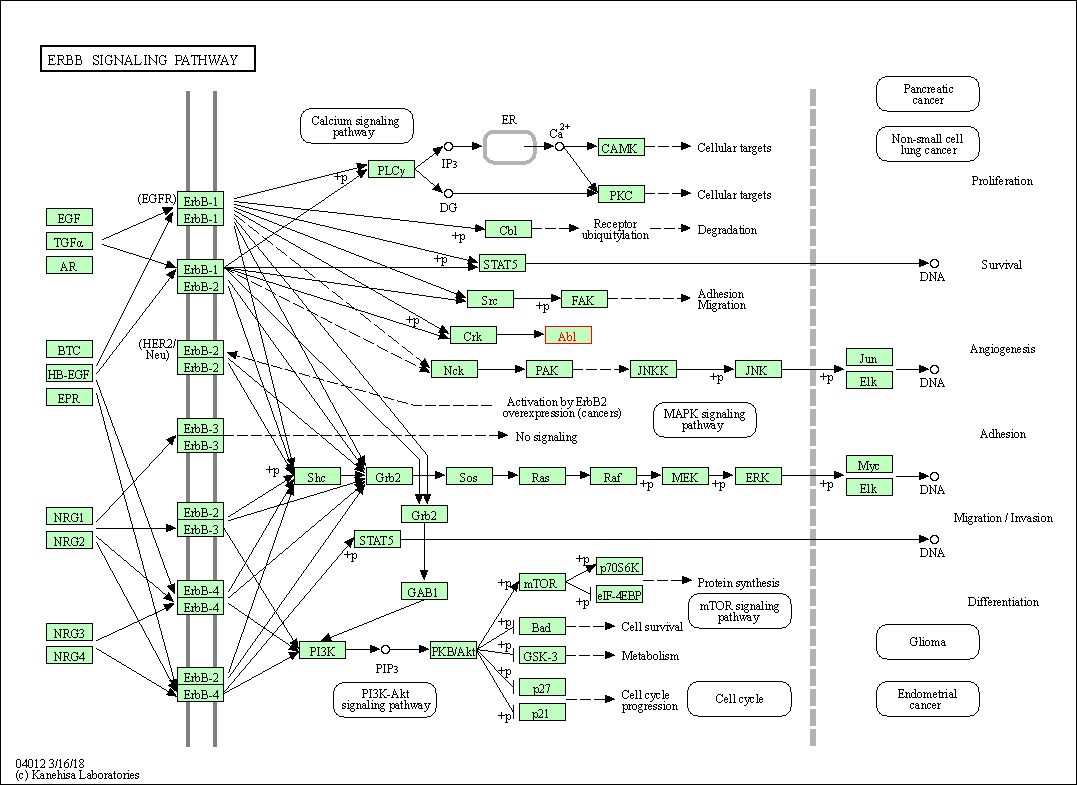
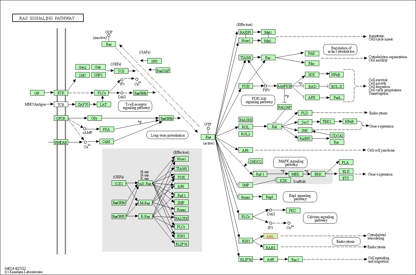
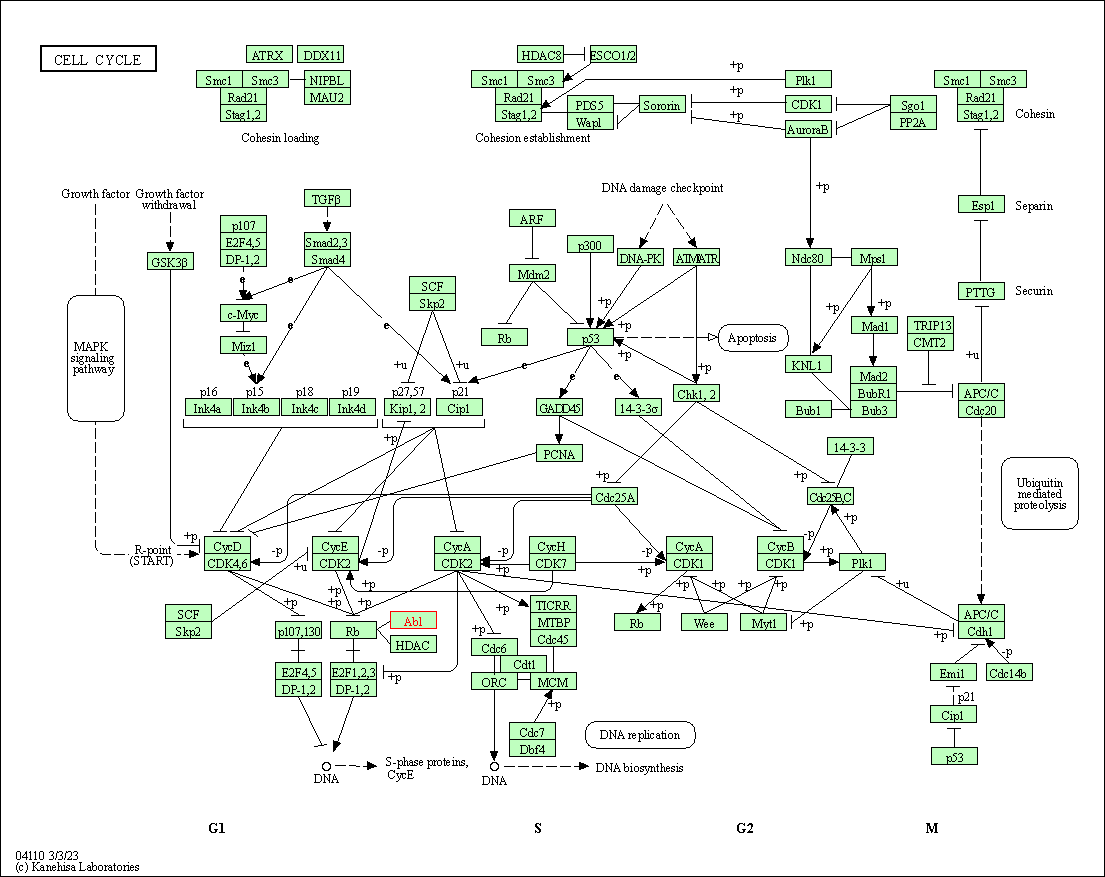
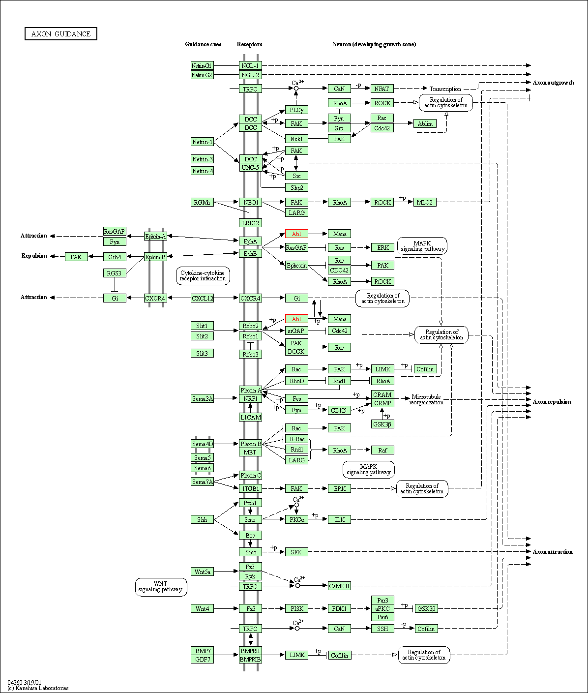
of target is determined by the life-essential pathways provided on KEGG database. The target-affiliated pathways were defined based on the following two criteria (a) the pathways of the studied target should be life-essential for both healthy individuals and patients, and (b) the studied target should occupy an upstream position in the pathways and therefore had the ability to regulate biological function.
Targets involved in a fewer pathways have greater likelihood to be successfully developed, while those associated with more human pathways increase the chance of undesirable interferences with other human processes
(Pharmacol Rev, 58: 259-279, 2006).
Human Similarity Proteins
Human Pathway Affiliation
|
|
| KEGG Pathway | Pathway ID | Affiliated Target | Pathway Map |
|---|---|---|---|
| ErbB signaling pathway | hsa04012 | Affiliated Target |
|
| Class: Environmental Information Processing => Signal transduction | Pathway Hierarchy | ||
| Ras signaling pathway | hsa04014 | Affiliated Target |
|
| Class: Environmental Information Processing => Signal transduction | Pathway Hierarchy | ||
| Cell cycle | hsa04110 | Affiliated Target |
|
| Class: Cellular Processes => Cell growth and death | Pathway Hierarchy | ||
| Axon guidance | hsa04360 | Affiliated Target |
|
| Class: Organismal Systems => Development and regeneration | Pathway Hierarchy | ||
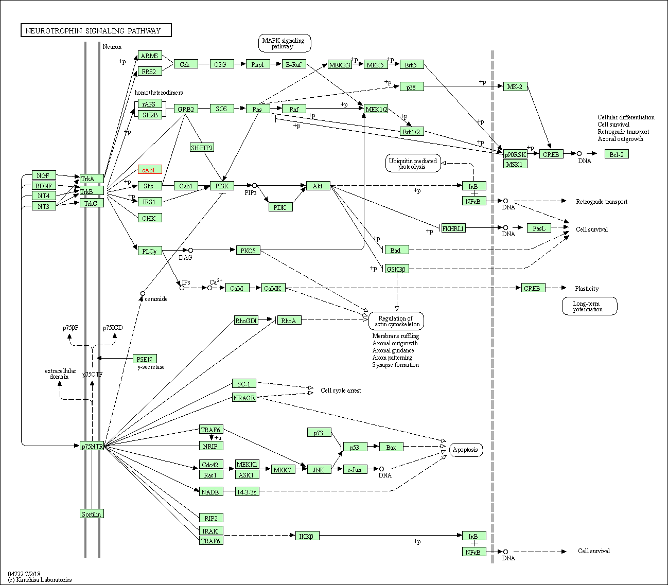
| Neurotrophin signaling pathway | hsa04722 | Affiliated Target |
|
| Class: Organismal Systems => Nervous system | Pathway Hierarchy | ||
| References | Top | |||||
|---|---|---|---|---|---|---|
| REF 1 | Bcr-Abl tyrosine kinase inhibitors: a patent review.Expert Opin Ther Pat. 2015 Apr;25(4):397-412. | |||||
| REF 2 | Axitinib effectively inhibits BCR-ABL1(T315I) with a distinct binding conformation. Nature. 2015 Mar 5;519(7541):102-5. | |||||
| REF 3 | Crystal structure of the T315I Abl mutant in complex with the aurora kinases inhibitor PHA-739358. Cancer Res. 2007 Sep 1;67(17):7987-90. | |||||
If You Find Any Error in Data or Bug in Web Service, Please Kindly Report It to Dr. Zhou and Dr. Zhang.

