Target Information
| Target General Information | Top | |||||
|---|---|---|---|---|---|---|
| Target ID |
T83009
(Former ID: TTDI03005)
|
|||||
| Target Name |
Tyrosine-protein kinase ABL2 (ABL2)
|
|||||
| Synonyms |
Tyrosine-protein kinase ARG; Abelson-related gene protein; Abelson tyrosine-protein kinase 2; Abelson murine leukemia viral oncogene homolog 2; ARG; ABLL
Click to Show/Hide
|
|||||
| Gene Name |
ABL2
|
|||||
| Target Type |
Literature-reported target
|
[1] | ||||
| Function |
Coordinates actin remodeling through tyrosine phosphorylation of proteins controlling cytoskeleton dynamics like MYH10 (involved in movement); CTTN (involved in signaling); or TUBA1 and TUBB (microtubule subunits). Binds directly F-actin and regulates actin cytoskeletal structure through its F-actin-bundling activity. Involved in the regulation of cell adhesion and motility through phosphorylation of key regulators of these processes such as CRK, CRKL, DOK1 or ARHGAP35. Adhesion-dependent phosphorylation of ARHGAP35 promotes its association with RASA1, resulting in recruitment of ARHGAP35 to the cell periphery where it inhibits RHO. Phosphorylates multiple receptor tyrosine kinases like PDGFRB and other substrates which are involved in endocytosis regulation such as RIN1. In brain, may regulate neurotransmission by phosphorylating proteins at the synapse. ABL2 acts also as a regulator of multiple pathological signaling cascades during infection. Pathogens can highjack ABL2 kinase signaling to reorganize the host actin cytoskeleton for multiple purposes, like facilitating intracellular movement and host cell exit. Finally, functions as its own regulator through autocatalytic activity as well as through phosphorylation of its inhibitor, ABI1. Non-receptor tyrosine-protein kinase that plays an ABL1-overlapping role in key processes linked to cell growth and survival such as cytoskeleton remodeling in response to extracellular stimuli, cell motility and adhesion and receptor endocytosis.
Click to Show/Hide
|
|||||
| BioChemical Class |
Kinase
|
|||||
| UniProt ID | ||||||
| EC Number |
EC 2.7.10.2
|
|||||
| Sequence |
MGQQVGRVGEAPGLQQPQPRGIRGSSAARPSGRRRDPAGRTTETGFNIFTQHDHFASCVE
DGFEGDKTGGSSPEALHRPYGCDVEPQALNEAIRWSSKENLLGATESDPNLFVALYDFVA SGDNTLSITKGEKLRVLGYNQNGEWSEVRSKNGQGWVPSNYITPVNSLEKHSWYHGPVSR SAAEYLLSSLINGSFLVRESESSPGQLSISLRYEGRVYHYRINTTADGKVYVTAESRFST LAELVHHHSTVADGLVTTLHYPAPKCNKPTVYGVSPIHDKWEMERTDITMKHKLGGGQYG EVYVGVWKKYSLTVAVKTLKEDTMEVEEFLKEAAVMKEIKHPNLVQLLGVCTLEPPFYIV TEYMPYGNLLDYLRECNREEVTAVVLLYMATQISSAMEYLEKKNFIHRDLAARNCLVGEN HVVKVADFGLSRLMTGDTYTAHAGAKFPIKWTAPESLAYNTFSIKSDVWAFGVLLWEIAT YGMSPYPGIDLSQVYDLLEKGYRMEQPEGCPPKVYELMRACWKWSPADRPSFAETHQAFE TMFHDSSISEEVAEELGRAASSSSVVPYLPRLPILPSKTRTLKKQVENKENIEGAQDATE NSASSLAPGFIRGAQASSGSPALPRKQRDKSPSSLLEDAKETCFTRDRKGGFFSSFMKKR NAPTPPKRSSSFREMENQPHKKYELTGNFSSVASLQHADGFSFTPAQQEANLVPPKCYGG SFAQRNLCNDDGGGGGGSGTAGGGWSGITGFFTPRLIKKTLGLRAGKPTASDDTSKPFPR SNSTSSMSSGLPEQDRMAMTLPRNCQRSKLQLERTVSTSSQPEENVDRANDMLPKKSEES AAPSRERPKAKLLPRGATALPLRTPSGDLAITEKDPPGVGVAGVAAAPKGKEKNGGARLG MAGVPEDGEQPGWPSPAKAAPVLPTTHNHKVPVLISPTLKHTPADVQLIGTDSQGNKFKL LSEHQVTSSGDKDRPRRVKPKCAPPPPPVMRLLQHPSICSDPTEEPTALTAGQSTSETQE GGKKAALGAVPISGKAGRPVMPPPQVPLPTSSISPAKMANGTAGTKVALRKTKQAAEKIS ADKISKEALLECADLLSSALTEPVPNSQLVDTGHQLLDYCSGYVDCIPQTRNKFAFREAV SKLELSLQELQVSSAAAGVPGTNPVLNNLLSCVQEISDVVQR Click to Show/Hide
|
|||||
| 3D Structure | Click to Show 3D Structure of This Target | AlphaFold | ||||
| Cell-based Target Expression Variations | Top | |||||
|---|---|---|---|---|---|---|
| Cell-based Target Expression Variations | ||||||
| Drug Binding Sites of Target | Top | |||||
|---|---|---|---|---|---|---|
| Ligand Name: Imatinib | Ligand Info | |||||
| Structure Description | The crystal structure of human ABL2 in complex with GLEEVEC | PDB:3GVU | ||||
| Method | X-ray diffraction | Resolution | 2.05 Å | Mutation | No | [2] |
| PDB Sequence |
GTENLYFQSM
278 DKWEMERTDI288 TMKHKLGGGQ298 YGEVYVGVWK308 KYSLTVAVKT318 LKEDTMEVEE 328 FLKEAAVMKE338 IKHPNLVQLL348 GVCTLEPPFY358 IVTEYMPYGN368 LLDYLRECNR 378 EEVTAVVLLY388 MATQISSAME398 YLEKKNFIHR408 DLAARNCLVG418 ENHVVKVADF 428 GLSRGDTYTA441 KFPIKWTAPE455 SLAYNTFSIK465 SDVWAFGVLL475 WEIATYGMSP 485 YPGIDLSQVY495 DLLEKGYRME505 QPEGCPPKVY515 ELMRACWKWS525 PADRPSFAET 535 HQAFETMFHD545 S
|
|||||
|
|
LEU294
3.911
TYR299
3.712
VAL302
3.662
ALA315
3.595
VAL316
3.953
LYS317
3.503
GLU332
2.911
VAL335
3.910
MET336
3.372
ILE339
3.745
LEU344
4.887
VAL345
3.335
ILE359
3.684
THR361
3.050
GLU362
4.375
TYR363
3.622
MET364
2.889
GLY367
4.055
ARG378
4.948
ALA383
3.006
VAL384
3.450
LEU386
3.708
LEU387
3.635
PHE405
4.224
ILE406
2.745
HIS407
3.204
ARG408
4.442
LEU416
3.779
VAL425
4.791
ALA426
3.409
ASP427
2.838
PHE428
3.376
LEU475
4.692
ILE478
4.967
ALA479
3.715
TYR481
3.929
GLU508
3.583
GLY509
3.570
CYS510
4.417
PRO511
3.685
VAL514
3.486
MET542
3.764
PHE543
4.131
SER546
4.153
|
|||||
| Ligand Name: VX-680 | Ligand Info | |||||
| Structure Description | HUMAN ABL2 IN COMPLEX WITH AURORA KINASE INHIBITOR VX-680 | PDB:2XYN | ||||
| Method | X-ray diffraction | Resolution | 2.81 Å | Mutation | No | [3] |
| PDB Sequence |
KWEMERTDIT
289 MKHKLGGGQY299 GEVYVGVWKK309 YSLTVAVKTL319 DTMEVEEFLK331 EAAVMKEIKH 341 PNLVQLLGVC351 TLEPPFYIVT361 EYMPYGNLLD371 YLRECNREEV381 TAVVLLYMAT 391 QISSAMEYLE401 KKNFIHRDLA411 ARNCLVGENH421 VVKVADFGLS431 RLMTGDTYTA 441 HAGAKFPIKW451 TAPESLAYNT461 FSIKSDVWAF471 GVLLWEIATY481 GMSPYPGIDL 491 SQVYDLLEKG501 YRMEQPEGCP511 PKVYELMRAC521 WKWSPADRPS531 FAETHQAFET 541 MFHD
|
|||||
|
|
LEU294
3.724
GLY295
4.337
TYR299
3.556
VAL302
3.204
ALA315
3.584
LYS317
3.689
GLU332
4.354
MET336
3.782
VAL345
4.116
ILE359
4.255
THR361
3.345
|
|||||
| Click to View More Binding Site Information of This Target with Different Ligands | ||||||
| Different Human System Profiles of Target | Top |
|---|---|
|
Human Similarity Proteins
of target is determined by comparing the sequence similarity of all human proteins with the target based on BLAST. The similarity proteins for a target are defined as the proteins with E-value < 0.005 and outside the protein families of the target.
A target that has fewer human similarity proteins outside its family is commonly regarded to possess a greater capacity to avoid undesired interactions and thus increase the possibility of finding successful drugs
(Brief Bioinform, 21: 649-662, 2020).
Human Tissue Distribution
of target is determined from a proteomics study that quantified more than 12,000 genes across 32 normal human tissues. Tissue Specificity (TS) score was used to define the enrichment of target across tissues.
The distribution of targets among different tissues or organs need to be taken into consideration when assessing the target druggability, as it is generally accepted that the wider the target distribution, the greater the concern over potential adverse effects
(Nat Rev Drug Discov, 20: 64-81, 2021).
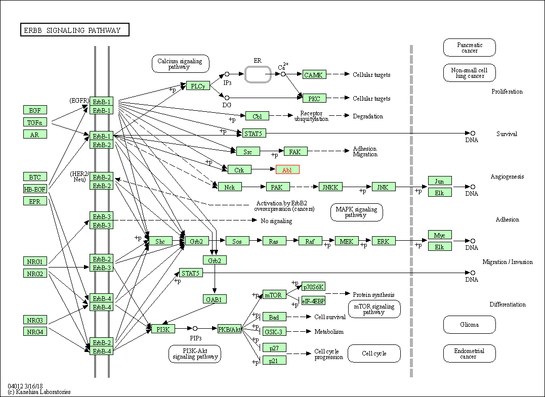
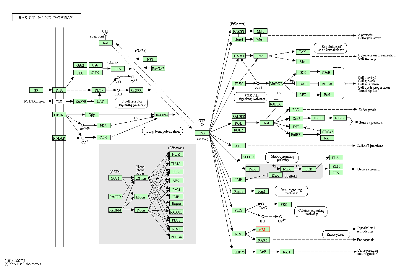
Human Pathway Affiliation
of target is determined by the life-essential pathways provided on KEGG database. The target-affiliated pathways were defined based on the following two criteria (a) the pathways of the studied target should be life-essential for both healthy individuals and patients, and (b) the studied target should occupy an upstream position in the pathways and therefore had the ability to regulate biological function.
Targets involved in a fewer pathways have greater likelihood to be successfully developed, while those associated with more human pathways increase the chance of undesirable interferences with other human processes
(Pharmacol Rev, 58: 259-279, 2006).
Biological Network Descriptors
of target is determined based on a human protein-protein interactions (PPI) network consisting of 9,309 proteins and 52,713 PPIs, which were with a high confidence score of ≥ 0.95 collected from STRING database.
The network properties of targets based on protein-protein interactions (PPIs) have been widely adopted for the assessment of target’s druggability. Proteins with high node degree tend to have a high impact on network function through multiple interactions, while proteins with high betweenness centrality are regarded to be central for communication in interaction networks and regulate the flow of signaling information
(Front Pharmacol, 9, 1245, 2018;
Curr Opin Struct Biol. 44:134-142, 2017).
Human Similarity Proteins
Human Tissue Distribution
Human Pathway Affiliation
Biological Network Descriptors
|
|
|
Note:
If a protein has TS (tissue specficity) scores at least in one tissue >= 2.5, this protein is called tissue-enriched (including tissue-enriched-but-not-specific and tissue-specific). In the plots, the vertical lines are at thresholds 2.5 and 4.
|


| KEGG Pathway | Pathway ID | Affiliated Target | Pathway Map |
|---|---|---|---|
| ErbB signaling pathway | hsa04012 | Affiliated Target |

|
| Class: Environmental Information Processing => Signal transduction | Pathway Hierarchy | ||
| Ras signaling pathway | hsa04014 | Affiliated Target |

|
| Class: Environmental Information Processing => Signal transduction | Pathway Hierarchy | ||
| Degree | 5 | Degree centrality | 5.37E-04 | Betweenness centrality | 4.91E-05 |
|---|---|---|---|---|---|
| Closeness centrality | 2.05E-01 | Radiality | 1.36E+01 | Clustering coefficient | 1.00E-01 |
| Neighborhood connectivity | 3.02E+01 | Topological coefficient | 2.19E-01 | Eccentricity | 13 |
| Download | Click to Download the Full PPI Network of This Target | ||||
| Chemical Structure based Activity Landscape of Target | Top |
|---|---|
| Target Poor or Non Binders | Top | |||||
|---|---|---|---|---|---|---|
| Target Poor or Non Binders | ||||||
| Target Regulators | Top | |||||
|---|---|---|---|---|---|---|
| Target-regulating microRNAs | ||||||
| Target-interacting Proteins | ||||||
| References | Top | |||||
|---|---|---|---|---|---|---|
| REF 1 | Rapid discovery of a novel series of Abl kinase inhibitors by application of an integrated microfluidic synthesis and screening platform. J Med Chem. 2013 Apr 11;56(7):3033-47. | |||||
| REF 2 | The crystal structure of human ABL2 in complex with GLEEVEC | |||||
| REF 3 | Crystal structures of ABL-related gene (ABL2) in complex with imatinib, tozasertib (VX-680), and a type I inhibitor of the triazole carbothioamide class. J Med Chem. 2011 Apr 14;54(7):2359-67. | |||||
If You Find Any Error in Data or Bug in Web Service, Please Kindly Report It to Dr. Zhou and Dr. Zhang.

