Target Information
| Target General Information | Top | |||||
|---|---|---|---|---|---|---|
| Target ID |
T35799
(Former ID: TTDI02598)
|
|||||
| Target Name |
DNA mismatch repair protein Mlh1 (MLH1)
|
|||||
| Synonyms |
MutL protein homolog 1; COCA2
Click to Show/Hide
|
|||||
| Gene Name |
MLH1
|
|||||
| Target Type |
Literature-reported target
|
[1] | ||||
| Function |
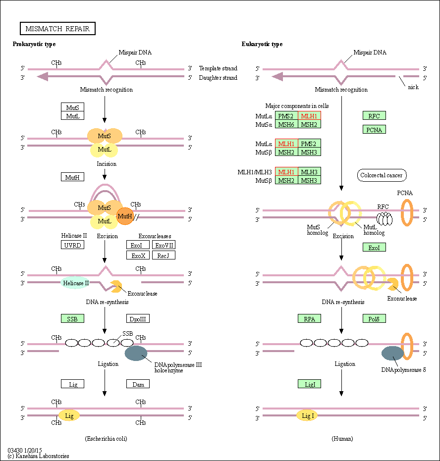
DNA repair is initiated by MutS alpha (MSH2-MSH6) or MutS beta (MSH2-MSH3) binding to a dsDNA mismatch, then MutL alpha is recruited to the heteroduplex. Assembly of the MutL-MutS-heteroduplex ternary complex in presence of RFC and PCNA is sufficient to activate endonuclease activity of PMS2. It introduces single-strand breaks near the mismatch and thus generates new entry points for the exonuclease EXO1 to degrade the strand containing the mismatch. DNA methylation would prevent cleavage and therefore assure that only the newly mutated DNA strand is going to be corrected. MutL alpha (MLH1-PMS2) interacts physically with the clamp loader subunits of DNA polymerase III, suggesting that it may play a role to recruit the DNA polymerase III to the site of the MMR. Also implicated in DNA damage signaling, a process which induces cell cycle arrest and can lead to apoptosis in case of major DNA damages. Heterodimerizes with MLH3 to form MutL gamma which plays a role in meiosis. Heterodimerizes with PMS2 to form MutL alpha, a component of the post-replicative DNA mismatch repair system (MMR).
Click to Show/Hide
|
|||||
| UniProt ID | ||||||
| Sequence |
MSFVAGVIRRLDETVVNRIAAGEVIQRPANAIKEMIENCLDAKSTSIQVIVKEGGLKLIQ
IQDNGTGIRKEDLDIVCERFTTSKLQSFEDLASISTYGFRGEALASISHVAHVTITTKTA DGKCAYRASYSDGKLKAPPKPCAGNQGTQITVEDLFYNIATRRKALKNPSEEYGKILEVV GRYSVHNAGISFSVKKQGETVADVRTLPNASTVDNIRSIFGNAVSRELIEIGCEDKTLAF KMNGYISNANYSVKKCIFLLFINHRLVESTSLRKAIETVYAAYLPKNTHPFLYLSLEISP QNVDVNVHPTKHEVHFLHEESILERVQQHIESKLLGSNSSRMYFTQTLLPGLAGPSGEMV KSTTSLTSSSTSGSSDKVYAHQMVRTDSREQKLDAFLQPLSKPLSSQPQAIVTEDKTDIS SGRARQQDEEMLELPAPAEVAAKNQSLEGDTTKGTSEMSEKRGPTSSNPRKRHREDSDVE MVEDDSRKEMTAACTPRRRIINLTSVLSLQEEINEQGHEVLREMLHNHSFVGCVNPQWAL AQHQTKLYLLNTTKLSEELFYQILIYDFANFGVLRLSEPAPLFDLAMLALDSPESGWTEE DGPKEGLAEYIVEFLKKKAEMLADYFSLEIDEEGNLIGLPLLIDNYVPPLEGLPIFILRL ATEVNWDEEKECFESLSKECAMFYSIRKQYISEESTLSGQQSEVPGSIPNSWKWTVEHIV YKALRSHILPPKHFTEDGNILQLANLPDLYKVFERC Click to Show/Hide
|
|||||
| 3D Structure | Click to Show 3D Structure of This Target | AlphaFold | ||||
| Cell-based Target Expression Variations | Top | |||||
|---|---|---|---|---|---|---|
| Cell-based Target Expression Variations | ||||||
| Drug Binding Sites of Target | Top | |||||
|---|---|---|---|---|---|---|
| Ligand Name: adenosine diphosphate | Ligand Info | |||||
| Structure Description | Crystal Structure of human MLH1 | PDB:4P7A | ||||
| Method | X-ray diffraction | Resolution | 2.30 Å | Mutation | No | [2] |
| PDB Sequence |
FVAGVIRRLD
12 ETVVNRIAAG22 EVIQRPANAI32 KEMIENCLDA42 KSTSIQVIVK52 EGGLKLIQIQ 62 DNGTGIRKED72 LDIVCERFTT82 SKLGFRGEAL104 ASISHVAHVT114 ITTKTADGKC 124 AYRASYSDGK134 LKAPPKPCAG144 NQGTQITVED154 LFYNIATRRK164 ALKNPSEEYG 174 KILEVVGRYS184 VHNAGISFSV194 KKQGETVADV204 RTLPNASTVD214 NIRSIFGNAV 224 SRELIEIGCE234 DKTLAFKMNG244 YISNANYSVK254 KCIFLLFINH264 RLVESTSLRK 274 AIETVYAAYL284 PKNTHPFLYL294 SLEISPESIL323 ERVQQHIESK333 LLG |
|||||
|
|
MET35
4.881
ASN38
3.053
CYS39
3.991
ASP41
4.592
ALA42
3.347
ASP63
2.923
THR66
4.682
GLY67
3.576
ILE68
3.391
VAL76
3.237
THR81
3.605
THR82
2.536
|
|||||
| Click to View More Binding Site Information of This Target with Different Ligands | ||||||
| Different Human System Profiles of Target | Top |
|---|---|
|
Human Similarity Proteins
of target is determined by comparing the sequence similarity of all human proteins with the target based on BLAST. The similarity proteins for a target are defined as the proteins with E-value < 0.005 and outside the protein families of the target.
A target that has fewer human similarity proteins outside its family is commonly regarded to possess a greater capacity to avoid undesired interactions and thus increase the possibility of finding successful drugs
(Brief Bioinform, 21: 649-662, 2020).
Human Tissue Distribution
of target is determined from a proteomics study that quantified more than 12,000 genes across 32 normal human tissues. Tissue Specificity (TS) score was used to define the enrichment of target across tissues.
The distribution of targets among different tissues or organs need to be taken into consideration when assessing the target druggability, as it is generally accepted that the wider the target distribution, the greater the concern over potential adverse effects
(Nat Rev Drug Discov, 20: 64-81, 2021).
Human Pathway Affiliation
of target is determined by the life-essential pathways provided on KEGG database. The target-affiliated pathways were defined based on the following two criteria (a) the pathways of the studied target should be life-essential for both healthy individuals and patients, and (b) the studied target should occupy an upstream position in the pathways and therefore had the ability to regulate biological function.
Targets involved in a fewer pathways have greater likelihood to be successfully developed, while those associated with more human pathways increase the chance of undesirable interferences with other human processes
(Pharmacol Rev, 58: 259-279, 2006).
Biological Network Descriptors
of target is determined based on a human protein-protein interactions (PPI) network consisting of 9,309 proteins and 52,713 PPIs, which were with a high confidence score of ≥ 0.95 collected from STRING database.
The network properties of targets based on protein-protein interactions (PPIs) have been widely adopted for the assessment of target’s druggability. Proteins with high node degree tend to have a high impact on network function through multiple interactions, while proteins with high betweenness centrality are regarded to be central for communication in interaction networks and regulate the flow of signaling information
(Front Pharmacol, 9, 1245, 2018;
Curr Opin Struct Biol. 44:134-142, 2017).
Human Similarity Proteins
Human Tissue Distribution
Human Pathway Affiliation
Biological Network Descriptors
|
|
|
There is no similarity protein (E value < 0.005) for this target
|
|
Note:
If a protein has TS (tissue specficity) scores at least in one tissue >= 2.5, this protein is called tissue-enriched (including tissue-enriched-but-not-specific and tissue-specific). In the plots, the vertical lines are at thresholds 2.5 and 4.
|


| KEGG Pathway | Pathway ID | Affiliated Target | Pathway Map |
|---|---|---|---|
| Mismatch repair | hsa03430 | Affiliated Target |

|
| Class: Genetic Information Processing => Replication and repair | Pathway Hierarchy | ||
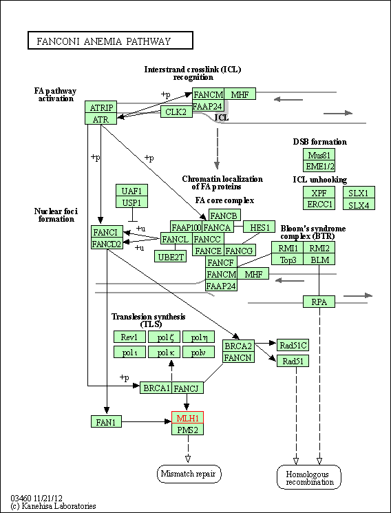
| Fanconi anemia pathway | hsa03460 | Affiliated Target |

|
| Class: Genetic Information Processing => Replication and repair | Pathway Hierarchy | ||
| Degree | 19 | Degree centrality | 2.04E-03 | Betweenness centrality | 7.88E-04 |
|---|---|---|---|---|---|
| Closeness centrality | 2.34E-01 | Radiality | 1.41E+01 | Clustering coefficient | 3.45E-01 |
| Neighborhood connectivity | 4.25E+01 | Topological coefficient | 1.02E-01 | Eccentricity | 12 |
| Download | Click to Download the Full PPI Network of This Target | ||||
| Target Regulators | Top | |||||
|---|---|---|---|---|---|---|
| Target-regulating microRNAs | ||||||
| Target-interacting Proteins | ||||||
| References | Top | |||||
|---|---|---|---|---|---|---|
| REF 1 | DNA mismatch repair gene MLH1 induces apoptosis in prostate cancer cells. Oncotarget. 2014 Nov 30;5(22):11297-307. | |||||
| REF 2 | Structure of the human MLH1 N-terminus: implications for predisposition to Lynch syndrome. Acta Crystallogr F Struct Biol Commun. 2015 Aug;71(Pt 8):981-5. | |||||
If You Find Any Error in Data or Bug in Web Service, Please Kindly Report It to Dr. Zhou and Dr. Zhang.

